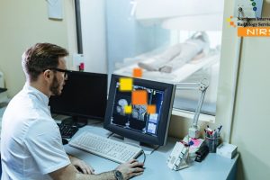
Best Guide to Breast Tumor Marker Clip Placement | NIRS
Breast cancer treatment has become more precise and patient-friendly thanks to advances in interventional radiology. One of the most effective tools for enhancing surgical precision and treatment planning is the breast tumor marker clip.
At NIRS (Northern Interventional Radiology Services), we perform image-guided, minimally invasive marker clip placements to ensure accurate tumor localization, safe procedures, and fast recovery. This step-by-step guide explains the process, benefits, and what patients can expect.
What Is a Breast Tumor Marker Clip?
A breast tumor marker clip is a tiny, biocompatible device placed inside or near a breast lesion. It serves multiple purposes:
- Accurate Localization: Helps surgeons locate small or non-palpable tumors for biopsy or surgery.
- Monitoring Tumor Changes: Tracks tumor response to chemotherapy or other therapies.
- Radiation Therapy Guidance: Provides a reference point for targeted radiation.
Marker clip placement is minimally invasive, usually performed under local anesthesia, and most patients can go home the same day.
Step 1: Pre-Procedure Consultation
Before the procedure, patients undergo a detailed consultation with an interventional radiologist. During this consultation:
- Review of medical history and imaging (ultrasound, mammography, MRI)
- Explanation of procedure benefits, risks, and alternatives
- Discussion of post-procedure care and recovery expectations
This step ensures patients are fully informed and comfortable with the procedure.
Step 2: Imaging and Tumor Localization
Accurate tumor localization is crucial for successful marker placement. Depending on the tumor location:
- Ultrasound guidance is used for visible or palpable tumors.
- Stereotactic mammography is used for small, non-palpable lesions.
- MRI guidance may be used for lesions only visible on MRI scans.
This imaging allows the radiologist to plan the exact path for marker placement, ensuring precision and safety.
Step 3: Local Anesthesia
The area surrounding the tumor is numbed with local anesthesia, making the procedure virtually painless. Unlike traditional surgery, general anesthesia is not required, reducing risks and recovery time.
Step 4: Marker Clip Insertion
Using a thin needle, the radiologist carefully inserts the marker clip into the tumor under real-time imaging guidance. The clip is tiny, usually less than 5mm, and stays securely in place without causing significant discomfort.
Step 5: Verification
After insertion, imaging (ultrasound or mammography) confirms correct clip placement. Accurate verification ensures that:
- Surgeons can easily locate the tumor during surgery
- Radiation therapy can be precisely targeted
- Follow-up monitoring of the tumor is reliable
Step 6: Post-Procedure Care
After marker placement:
- Mild bruising or discomfort at the insertion site is normal
- Most patients can return home the same day
- Light activities can resume immediately, but avoid heavy lifting for 24–48 hours
- Follow-up imaging may be scheduled to confirm marker stability
The minimally invasive nature of this procedure allows patients to resume daily life quickly.
Benefits of Marker Clip Placement
- Surgical Precision: Ensures complete tumor removal while sparing healthy tissue
- Minimally Invasive: Small puncture, minimal pain, low infection risk
- Supports Multiple Treatments: Useful for surgery, chemotherapy, and radiation
- Faster Recovery: Outpatient procedure with minimal downtime
- Better Cosmetic Results: Preserves breast tissue and appearance
Who Can Benefit From This Procedure?
- Women with small, hard-to-locate tumors
- Patients undergoing neoadjuvant chemotherapy
- Individuals preparing for breast-conserving surgery or lumpectomy
- Patients requiring precision-targeted radiation therapy
- Anyone seeking minimally invasive, safe breast care
At NIRS, we provide personalized guidance to ensure every patient understands the procedure, risks, and recovery process.
Real-Life Impact
| Condition | Traditional Surgery | Marker Clip Placement at NIRS |
|---|---|---|
| Small or non-palpable tumor | Large incision, guesswork | Precise localization via clip |
| Tumor shrinking during chemo | Hard to locate | Marker maintains exact tumor location |
| Radiation therapy planning | Broad targeting | Targeted therapy using clip reference |
| Multiple lesions | Surgical uncertainty | Accurate mapping and excision of all tumors |
Marker clips ensure precision, safety, and better overall outcomes.
Why Choose NIRS for Marker Clip Placement?
✔ Experienced interventional radiologists trained in advanced breast imaging
✔ State-of-the-art imaging guidance for ultrasound, mammography, and stereotactic procedures
✔ Minimally invasive, outpatient approach
✔ Same-day discharge and rapid recovery
✔ Trusted across Northern Pakistan and Azad Kashmir
At NIRS, precision, safety, and patient comfort are top priorities.
Final Thoughts
Breast tumor marker clip placement is a revolutionary tool in modern breast cancer care. This step-by-step guide illustrates how the procedure:
- Improves surgical accuracy
- Supports minimally invasive treatment
- Enhances cosmetic and medical outcomes
- Reduces recovery time
At NIRS, we combine advanced interventional radiology with personalized patient care to make marker clip placement safe, precise, and stress-free.
For patients seeking precision, minimally invasive treatment, and optimal breast cancer care, NIRS is the trusted choice in Northern Pakistan.