Early Cancer Detection With Advanced and Best Imaging | NIRS
Importance of Early Detection
Early cancer detection is one of the most powerful tools in modern medicine. Catching cancer in its early stages dramatically improves treatment success, survival rates, and quality of life. When cancer is diagnosed before it spreads, patients often require less aggressive treatment, experience fewer side effects, and recover faster.
At Northern Interventional Radiology Services (NIRS), we prioritize proactive health screening through high-resolution imaging techniques such as CT scans, MRI, and ultrasound-guided evaluations. These advanced diagnostic tools help our specialists detect abnormalities even before symptoms arise, giving patients the advantage of early intervention.
Timely diagnosis leads to timely care. Whether it’s identifying lung nodules, breast lesions, or liver tumors, NIRS ensures precise, minimally invasive diagnostics backed by state-of-the-art technology. With our experienced interventional radiologists and modern imaging systems, patients can trust NIRS to lead them toward better health outcomes through early detection.
Remember, early cancer detection can mean the difference between a simple procedure and a complex treatment journey. Choose NIRS for advanced screening and diagnostic excellence.
🧠 Understanding Advanced Medical Imaging
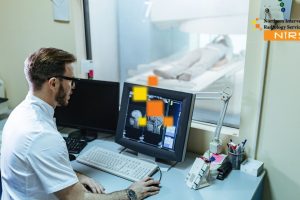
Medical imaging is one of the most transformative innovations in healthcare. It allows doctors to visualize internal organs, tissues, and abnormalities with incredible precision—without the need for invasive procedures. At Northern Interventional Radiology Services (NIRS), advanced imaging plays a pivotal role in diagnosing complex diseases and enabling early cancer detection, improving patient outcomes drastically.
What is Advanced Medical Imaging?
Advanced medical imaging refers to sophisticated technologies used to capture detailed images of the human body. These include:
- MRI (Magnetic Resonance Imaging)
- CT Scan (Computed Tomography)
- Ultrasound Imaging
- PET Scans (Positron Emission Tomography)
- Digital X-rays
These modalities help identify tumors, internal bleeding, organ abnormalities, and other serious conditions at very early stages.
Role in Early Cancer Detection
One of the most critical applications of medical imaging is in early cancer detection. When cancer is identified early—before symptoms even arise—it allows for:
- Less aggressive treatment options
- Higher survival rates
- Reduced long-term complications
- Lower healthcare costs
At NIRS, high-resolution imaging tools are used to detect subtle changes in tissues that could indicate the early onset of cancer.
How It Enhances Diagnostic Accuracy
Medical imaging is not only about seeing what’s wrong—it’s about seeing it clearly and early. With technologies like contrast-enhanced CT or multiparametric MRI, radiologists at NIRS can distinguish between benign and malignant masses, reducing unnecessary biopsies or exploratory surgeries.
The Technology Behind It
Behind every clear image lies powerful hardware and software:
- High-frequency ultrasound probes
- AI-powered imaging software
- 3D reconstruction tools
- Portable and mobile imaging units
These tools work together to create precise and real-time visuals, helping radiologists make better clinical decisions.
💡 How Early Detection Improves Cancer Survival Rates
Early cancer detection plays a transformative role in saving lives. At Northern Interventional Radiology Services (NIRS), we emphasize the importance of spotting cancer at its earliest stages to increase the chances of successful treatment and long-term survival.
Survival Rates Are Significantly Higher
When cancer is detected early—before it spreads to other organs—the chances of survival are significantly higher. According to global health data:
- Breast cancer detected in Stage I has a 5-year survival rate of nearly 100%.
- Colon cancer found early has a survival rate of over 90%.
- Lung cancer, which is often diagnosed late, has dramatically better outcomes if caught in Stage I.
These statistics prove that timing is critical in treatment planning.
More Treatment Options Available
Early-stage cancers often require less aggressive treatment. With early cancer detection, patients may:
- Avoid chemotherapy or undergo limited radiation.
- Opt for minimally invasive surgeries.
- Experience fewer side effects and quicker recovery.
At NIRS, advanced imaging technologies help doctors identify cancer when it’s small and manageable—unlocking more effective and patient-friendly treatment strategies.
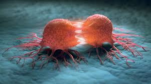
Reduced Risk of Metastasis
One of the greatest threats in cancer is metastasis—the spread of cancer to other parts of the body. Early detection limits this risk, allowing treatment to remain localized and more successful.
Imaging techniques like PET-CT and MRI offered at NIRS help track cancerous growths with precision before they metastasize, preserving the patient’s overall health and organ function.It is also helpful at Early cancer detection.
Lower Healthcare Costs and Emotional Strain
Early diagnosis often results in shorter treatment durations and fewer complications, which can reduce the financial and emotional burden on both patients and families.
Preventive imaging and screening services at NIRS aim not only to detect disease but also to support patients with less disruptive treatment pathways.
🖼️ Imaging Technologies Used in Cancer Diagnosis
Accurate and timely cancer diagnosis depends heavily on advanced imaging technologies. At Northern Interventional Radiology Services (NIRS), we utilize a wide range of cutting-edge imaging tools that help detect tumors, assess their progression, and guide treatment strategies.
1. Computed Tomography (CT Scan)
A CT scan combines X-ray images taken from multiple angles to create cross-sectional views of the body. It is especially useful for:
- Detecting lung, liver, pancreatic, and abdominal cancers
- Identifying tumor size, shape, and exact location
- Evaluating the spread of cancer to nearby tissues
CT scans are fast and non-invasive, making them a routine part of Early cancer detection.
2. Magnetic Resonance Imaging (MRI)
MRI uses powerful magnets and radio waves to produce detailed images of organs and tissues. It’s especially effective for:
- Brain and spinal cord tumors
- Breast and pelvic cancers
- Evaluating soft tissue contrast without radiation exposure
At NIRS, MRI is a core part of our diagnostic protocol, particularly when radiation-free imaging is preferred.
3. Positron Emission Tomography (PET Scan)
PET scans use a small amount of radioactive tracer to detect metabolic activity. Cancer cells typically absorb this tracer more than normal cells, revealing:
- Active cancer regions
- Metastasis or recurrence
- Response to treatment over time
PET-CT (combined PET and CT) enhances diagnostic accuracy and is commonly used at NIRS for staging and monitoring therapy effectiveness.
4. Ultrasound Imaging
Ultrasound uses high-frequency sound waves to create real-time images, particularly useful for:
- Breast, liver, and pelvic area assessments
- Guiding biopsies or needle aspirations
- Evaluating blood flow in suspicious areas
Ultrasound is safe, cost-effective, and ideal for initial screening, especially in younger patients or those with dense tissues.
5. X-Rays
While basic, X-rays remain a first-line imaging technique for detecting abnormalities in bones and lungs. They help:
- Identify fractures caused by bone tumors
- Detect early signs of lung nodules
- Guide physicians toward further tests when something unusual appears
X-rays are often the starting point before advancing to more detailed imaging methods.
6. Mammography
Mammography is a specialized type of X-ray specifically for breast cancer screening. It can detect:
- Calcifications and masses
- Structural distortions in breast tissue
- Early-stage cancer before symptoms appear
At NIRS, mammography services are offered as part of our routine breast cancer screening programs.
7. Fluoroscopy and Image-Guided Biopsy
For confirming cancer after suspicious imaging findings, we use fluoroscopy or image-guided techniques to perform precise biopsies. These procedures ensure:
- Accurate tissue sampling
- Minimal invasiveness
- Improved diagnostic reliability
These imaging technologies collectively support the most comprehensive and personalized Early cancer detection approaches at Northern Interventional Radiology Services (NIRS). By choosing the right modality based on the suspected cancer type and location, we ensure early, accurate detection and optimized treatment planning.
👩⚕️ Role of Radiologists in Early Detection
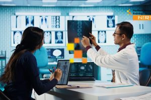
Radiologists play a pivotal role in the Early cancer detection and other critical diseases. Their expertise in interpreting medical imaging is essential to identifying abnormalities at a stage when treatment is most effective. At Northern Interventional Radiology Services (NIRS), our skilled radiologists are central to every patient’s diagnostic journey.
1. Expert Image Interpretation
Radiologists are trained to interpret complex imaging results from X-rays, CT scans, MRIs, PET scans, and ultrasounds. They can detect:
- Tiny lesions or suspicious masses
- Irregular tissue patterns
- Signs of early malignancy that may be missed by the untrained eye
Their insights are vital for accurate diagnosis and timely referral to oncologists or specialists.
2. Screening Program Participation
Radiologists actively participate in national and regional cancer screening programs, including:
- Mammography for breast cancer
- Low-dose CT for lung cancer
- Ultrasound for thyroid or abdominal cancers
Their role includes both reading the scans and guiding the development of screening protocols that prioritize early detection.
3. Guidance for Biopsies and Interventions
When a suspicious area is found, radiologists assist in image-guided biopsies, ensuring:
- Precise targeting of abnormal tissue
- Minimally invasive procedures
- Lower risk and faster recovery for patients
This accuracy increases the likelihood of an Early cancer detection.
4. Collaboration with Multidisciplinary Teams
Radiologists work closely with surgeons, oncologists, and primary care doctors. Their collaboration includes:
- Presenting imaging findings at tumor board meetings
- Recommending further diagnostic tests
- Helping design personalized treatment plans
This team-based approach ensures no early signs of cancer go unnoticed.= and for Early cancer detection.
5. Monitoring and Follow-Up
Even after initial diagnosis, radiologists remain involved by:
- Comparing past and current imaging to detect recurrence
- Monitoring treatment effectiveness
- Alerting teams to progression or regression of disease
Their ongoing assessments are key to successful long-term outcomes.
6. Patient Education and Safety
Radiologists also focus on educating patients and ensuring safety in imaging:
- Advising on low-dose imaging to minimize radiation exposure
- Explaining the necessity and results of imaging tests
- Supporting informed decisions on next steps
This adds trust and transparency to the care process.
Radiologists are often the first line of defense in identifying cancers before symptoms develop. At NIRS, their vigilance, precision, and deep understanding of imaging science form the backbone of early cancer detection—improving patient survival rates and enhancing quality of care.
🧬 Common Cancers Identified Through Imaging
Medical imaging plays a critical role in the early cancer detection process by identifying tumors and abnormalities before symptoms appear. At Northern Interventional Radiology Services (NIRS), advanced imaging techniques are used to detect a wide range of cancers, allowing for timely intervention and improved patient outcomes.
1. Breast Cancer
- Mammography is the most effective screening tool for early detection of breast cancer.
- It identifies small masses or microcalcifications that may indicate early malignancy.
- Ultrasound and MRI are used as complementary methods, especially in high-risk individuals or dense breast tissue.
2. Lung Cancer
- Low-dose CT (LDCT) scans are widely used to screen for lung cancer, especially in smokers or those with a history of smoking.
- LDCT helps identify small lung nodules before symptoms such as coughing or breathlessness begin.
3. Colorectal Cancer
- CT Colonography (Virtual Colonoscopy) allows non-invasive imaging of the colon and rectum.
- It detects polyps and tumors that could become cancerous if left untreated.
- This method is a less invasive alternative to conventional colonoscopy for screening.
4. Liver Cancer
- Ultrasound, CT scans, and MRI are often used to screen and monitor patients at risk of hepatocellular carcinoma (HCC).
- Imaging is crucial for detecting liver lesions in early, treatable stages.
5. Prostate Cancer
- MRI of the prostate can detect suspicious areas and guide biopsy decisions.
- It provides detailed images that help differentiate between benign and potentially malignant conditions.
6. Pancreatic Cancer
- CT and MRI scans are used to detect pancreatic tumors, which are often difficult to diagnose early due to vague symptoms.
- Imaging helps in both diagnosis and staging for surgical planning.
7. Thyroid Cancer
- Neck ultrasound is the first-line imaging technique for evaluating thyroid nodules.
- It can identify features suggestive of cancer and guide fine-needle aspiration biopsy.
8. Bladder and Kidney Cancers
- Ultrasound, CT urography, and MRI help detect tumors in the urinary tract.
- These imaging tools are essential for identifying masses, structural abnormalities, and blood in the urine source.
9. Brain Tumors
- MRI is the gold standard for brain imaging.
- It can detect tumors of the brain and spinal cord, even at small sizes, allowing for earlier neurosurgical planning or treatment.
10. Ovarian and Uterine Cancers
- Transvaginal ultrasound, MRI, and CT scans are commonly used for detection and staging.
- These tools help evaluate abnormal bleeding or pelvic masses.
At NIRS, early imaging detection of these cancers ensures that patients receive prompt care and have better chances of recovery. Our radiologists are trained in identifying the subtle signs of malignancy, helping to save lives through precision diagnostics for early cancer detection.
🔮 Future Trends in Cancer Imaging and Diagnostics
The field of cancer imaging is rapidly evolving, integrating advanced technologies and artificial intelligence to improve accuracy, speed, and patient outcomes. As diagnostic tools continue to innovate, early cancer detection is becoming more precise, less invasive, and more personalized.Which also helpful for Early cancer detection.
1. Artificial Intelligence (AI) and Machine Learning
AI is transforming how imaging is interpreted:
- Algorithms can analyze complex imaging data faster and more accurately than traditional methods.
- AI assists radiologists in detecting subtle changes that may indicate early cancer.
- Machine learning models improve over time, refining diagnostics with each scan they process.
2. Molecular Imaging
This next-gen technique allows visualization of biological processes at the cellular and molecular levels:
- Detects cancer activity before structural changes are visible.
- Combines imaging with biomarkers to monitor tumor response to therapy.
- Commonly used in PET scans with radiotracers targeting specific cancer types.
3. Hybrid Imaging Technologies
Integrating multiple imaging modalities for more comprehensive diagnostics:
- PET/CT and PET/MRI offer both anatomical and functional insights in one scan.
- Improves detection of small tumors and metastases.
- Helps in treatment planning and monitoring response to therapies.
4. Radiomics and Radiogenomics
Analyzing vast amounts of imaging data beyond the visible spectrum:
- Radiomics extracts quantitative features from scans to detect patterns linked to cancer.
- Radiogenomics links imaging features with genetic data, helping predict tumor behavior and treatment response.
- These technologies are paving the way for personalized medicine.
5. Liquid Biopsy Integration
Combining imaging with non-invasive blood tests:
- Liquid biopsies detect circulating tumor DNA (ctDNA) or cancer cells.
- Imaging guides the biopsy timing and confirms treatment response.
- This synergy enhances early diagnosis and reduces unnecessary interventions.
6. Portable and Point-of-Care Imaging
Developments in miniaturized imaging equipment:
- Enables early detection in rural or low-resource settings.
- Portable ultrasound, handheld MRI, and mobile CT devices are expanding global cancer care.
- Ideal for follow-up or screening without needing hospital visits.
7. 3D Imaging and Virtual Reality (VR)
Revolutionizing visualization and planning:
- 3D reconstructions help surgeons plan complex cancer surgeries.
- VR allows medical teams to navigate tumors digitally before actual procedures.
- Enhances patient understanding and engagement with their treatment plan.
8. Improved Contrast Agents
Next-generation contrast materials offer safer and more effective visualization:
- Designed to target tumors more specifically.
- Lower toxicity and improved image clarity.
- Particularly beneficial in detecting hidden or early-stage cancers.
9. Tele-radiology and Cloud-Based Diagnostics
Remote interpretation of imaging is reshaping global diagnostics:
- Enables access to expert radiologists across borders.
- Cloud platforms allow faster collaboration between oncologists, radiologists, and pathologists.
- Ideal for faster second opinions and continuity of care.
10. Focus on Early and Preventive Imaging
The future of cancer care emphasizes prevention:
- More widespread use of imaging in routine health checkups.
- AI-assisted tools for screening high-risk populations.
- Early-stage diagnosis improves survival rates and reduces treatment costs.
✅ Conclusion
Cancer imaging has revolutionized how we detect, diagnose, and treat cancer. From traditional imaging technologies like CT and MRI to cutting-edge advancements in AI, radiomics, and molecular imaging, these tools allow for earlier and more accurate early cancer detection. Radiologists play a pivotal role not only in interpreting these images but also in guiding biopsies, planning treatments, and monitoring outcomes. With cancers like breast, lung, prostate, and colorectal commonly identified through imaging, early and accurate diagnosis has never been more critical. This will make easy for early cancer detection.
Looking ahead, the future of cancer imaging is full of promise. Artificial intelligence, hybrid imaging, portable diagnostics, and integration with genetic profiling will pave the way for personalized cancer care. At Northern Interventional Radiology Services (NIRS), we are committed to staying at the forefront of these technologies to ensure patients receive the most accurate and least invasive diagnostic solutions for early cancer detection.
❓ Frequently Asked Questions (FAQs)
1. What is the role of imaging in cancer diagnosis?
Imaging helps detect tumors, determine their location and size, guide biopsies, and monitor treatment response.
2. Which imaging methods are most commonly used for cancer?
Common methods include MRI, CT scans, PET scans, ultrasound, and X-rays, depending on the type of cancer.
3. How do radiologists help in early cancer detection?
Radiologists interpret imaging studies and can spot abnormal changes before symptoms arise, leading to earlier diagnosis and treatment.
4. What cancers are most commonly detected through imaging?
Breast, lung, liver, prostate, and colorectal cancers are frequently identified through imaging techniques.
5. Are modern imaging methods safe?
Yes, most imaging methods are safe. Advanced technologies use low-dose radiation and non-invasive approaches, making them patient-friendly for early cancer detection.
6. Can AI replace radiologists in cancer diagnosis?
AI supports radiologists but does not replace them. It enhances accuracy and efficiency, but human expertise remains essential.
7. What is molecular imaging?
Molecular imaging shows biological activity at the molecular level, helping detect cancer early and monitor its response to treatment of early cancer detection.
8. What are hybrid imaging techniques?
These combine two imaging modalities (like PET/CT) to provide both structural and functional information for more accurate diagnosis and early cancer detection.
9. Is cancer imaging painful?
Most cancer imaging procedures are non-invasive and painless, though some may require contrast injections or mild preparation for best early cancer detection.
10. Where can I get advanced cancer imaging in Haripur?
You can consult Northern Interventional Radiology Services (NIRS) for advanced, affordable, and reliable early cancer detection solutions.
📞 Contact Us
If you or a loved one needs early cancer detection or imaging services, we are here to help. Reach out to Northern Interventional Radiology Services (NIRS) through the following channels:
- 📞 Phone: +92316 5212015
- 📧 Email: info@nirs.com.pk
- 🌐 Website: www.nirs.com.pk
- 🔵 Facebook: NIRS
- 🟣 Instagram: NIRS
Our team, including expert radiologists like Dr. Muhammad Ismail and Dr. Atif Nawaz, is dedicated to providing safe, accurate, and timely diagnostics to aid in early cancer detection.