Breakthrough Diagnostic Imaging for Early Disease Detection | NIRS
Introduction to Diagnostic Imaging
Diagnostic imaging is a cornerstone of modern medicine, providing detailed, non-invasive insight into the body’s internal structures. It plays a pivotal role in identifying, monitoring, and treating a wide range of health conditions. At NIRS (Northern Institute of Radiological Sciences) in Abbottabad, diagnostic imaging services are not just about scans—they’re about early detection, precise diagnosis, and patient-centric care.
Whether it’s through X-rays, CT scans, MRIs, ultrasound, or interventional radiology, NIRS offers advanced imaging techniques that aid physicians in making accurate clinical decisions. These technologies reduce the need for exploratory surgeries and allow for early disease detection, which significantly improves treatment outcomes.
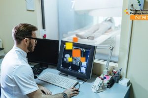
At NIRS, every diagnostic procedure is conducted using cutting-edge equipment under the supervision of expert radiologists and technologists. Their focus on safety, precision, and patient comfort ensures that individuals receive the highest standard of radiological care in the region.
What is Diagnostic Imaging?
Diagnostic imaging refers to a set of non-invasive techniques that allow medical professionals to view structures and processes inside the body. It plays an essential role in detecting diseases, assessing injuries, monitoring treatment progress, and guiding medical procedures. At NIRS, diagnostic imaging is performed with the latest technology to ensure high-resolution, precise visual representations of internal anatomy.
The main types of diagnostic imaging include X-rays, ultrasound, CT (Computed Tomography) scans, MRI (Magnetic Resonance Imaging), mammography, and nuclear medicine scans. These tools are vital for diagnosing conditions like fractures, tumors, infections, and cardiovascular diseases. At NIRS Abbottabad, patients receive tailored imaging solutions based on their symptoms and medical history, ensuring accurate diagnoses and optimized treatment plans.
By utilizing state-of-the-art equipment and following global radiological standards, NIRS ensures safe radiation doses, minimal discomfort, and quick results—making it a trusted center for diagnostic imaging in the region.
Historical Evolution of Imaging Technologies
The field of diagnostic imaging has undergone a remarkable transformation since its inception. It began in 1895 when Wilhelm Conrad Roentgen discovered X-rays, revolutionizing medical diagnostics by allowing doctors to see inside the human body without surgery. This single discovery laid the foundation for modern radiology.
In the decades that followed, the development of ultrasound in the 1950s, CT scans in the 1970s, and MRI in the 1980s expanded the capabilities of imaging significantly. Each innovation brought clearer images, enhanced safety, and broader diagnostic scope. By the 2000s, digital imaging and 3D reconstruction became mainstream, further advancing diagnostic precision.
Today, at NIRS, this rich history is reflected in their adoption of the most advanced diagnostic imaging technologies. From high-resolution MRI scanners to low-radiation CT systems, NIRS ensures patients benefit from decades of scientific progress. Their radiologists are trained in both the science and history of imaging, enabling them to apply a deep understanding of imaging modalities to every case.
By combining a historical foundation with modern-day innovation, NIRS has positioned itself as a leader in radiological excellence across Khyber Pakhtunkhwa.
Importance of Early Disease Detection
Early disease detection plays a pivotal role in modern healthcare. It allows medical professionals to identify illnesses at their initial stages—when they are most treatable and least damaging. At NIRS, early detection is made possible through the strategic use of advanced diagnostic imaging, which offers a clear and non-invasive window into the body’s internal systems.
By spotting abnormalities before they progress, NIRS diagnostic imaging supports proactive healthcare, reduces complications, and increases the chances of successful treatment. This approach is particularly valuable for life-threatening conditions like cancer, cardiovascular disease, neurological disorders, and internal injuries—many of which show no outward symptoms until they’ve advanced.
Why Early Detection Matters in Modern Medicine
In today’s rapidly advancing medical field, early detection is not just beneficial—it’s essential. Diseases like cancer, tuberculosis, and heart conditions often remain hidden during their early phases. Using diagnostic imaging technologies, such as CT, MRI, and ultrasound, NIRS empowers physicians to detect these diseases before they escalate.
With early detection:
- Treatment can start sooner, often leading to better prognosis.
- Patients may avoid invasive surgeries and high-risk interventions.
- Chronic conditions can be managed or even reversed in early stages.
- Healthcare costs are often significantly reduced.
At NIRS, these principles are applied daily. Their diagnostic imaging tools are calibrated to identify even the smallest abnormalities. This ensures no early warning signs are missed, supporting physicians in their clinical decisions.
Impact on Treatment Outcomes and Survival Rates
One of the most compelling reasons for early detection through diagnostic imaging is its direct impact on survival rates and treatment outcomes. When diseases are diagnosed at an early stage, the likelihood of full recovery increases dramatically. For example:
- Early-stage cancer patients often have a 90%+ survival rate, compared to much lower rates in later stages.
- Early identification of stroke signs via MRI or CT scans allows doctors at NIRS to intervene swiftly, reducing permanent neurological damage.
- Cardiovascular issues caught through echocardiograms or angiography can be treated before resulting in heart attacks or heart failure.
At NIRS, each imaging report is handled with care and urgency. Their specialists ensure patients are not only diagnosed early but are also connected with the appropriate care pathways. By prioritizing early disease detection, NIRS improves outcomes and helps patients reclaim healthier lives faster.
How Diagnostic Imaging Aids in Early Detection
Diagnostic imaging is the cornerstone of modern medical diagnosis, especially when it comes to early disease detection. Through non-invasive technologies, medical professionals can visualize internal body structures and spot abnormalities long before symptoms arise. At NIRS (Northern Institute of Radiological Sciences), cutting-edge diagnostic imaging is used to uncover hidden threats—often the difference between a manageable condition and a life-threatening illness.
With modalities like MRI, CT scan, ultrasound, and X-ray, NIRS diagnostic imaging offers physicians an unparalleled ability to identify disease at its root. These insights empower both patients and doctors to take action early, often before irreversible damage occurs.
Role in Identifying Hidden or Asymptomatic Conditions
One of the most important benefits of diagnostic imaging is its ability to detect conditions that show no visible or physical symptoms. These are often referred to as asymptomatic diseases, and they can include early-stage cancers, internal bleeding, brain tumors, bone infections, and even congenital anomalies.
At NIRS, diagnostic imaging is used to:
- Identify lung nodules or lesions before respiratory symptoms develop.
- Detect brain aneurysms or clots before they cause a stroke.
- Reveal silent kidney stones or gallbladder issues.
- Monitor changes in breast tissue through mammography before any lump is felt.
By diagnosing these silent threats early, NIRS imaging experts help patients avoid emergency situations, reduce complications, and plan proactive treatment strategies. This approach improves quality of life and reduces the emotional and financial burdens that come with late-stage diagnoses.
Imaging Techniques That Detect Disease Before Symptoms Appear
Not all diseases announce themselves with pain, swelling, or fatigue. In fact, many life-threatening illnesses progress quietly. Diagnostic imaging techniques used at NIRS are designed specifically to detect disease before symptoms appear—a revolutionary advantage in preventive healthcare.
Some of the most effective early detection imaging techniques include:
- Magnetic Resonance Imaging (MRI): Ideal for soft tissues, brain structures, and joint assessments. Helps detect tumors, early-stage MS, and neurodegenerative diseases.
- Computed Tomography (CT): Provides detailed cross-sectional views of the body. CT is highly effective in catching small lung nodules, liver issues, and internal bleeding.
- Ultrasound: Non-invasive and radiation-free. Often used in obstetrics, abdominal scans, and vascular health. Detects cysts, fibroids, and organ enlargement early.
- Mammography: Specialized X-ray used for early breast cancer detection.
- Bone Densitometry (DEXA): Detects low bone density before fractures occur, aiding in osteoporosis management.
At NIRS, these technologies are operated by skilled radiologists and technologists who ensure accuracy, precision, and timely reporting. Whether for a routine check-up or a specific concern, NIRS diagnostic imaging is equipped to deliver early insights that can change lives.
Common Types of Diagnostic Imaging
At the core of modern healthcare lies the ability to see what the naked eye cannot. Diagnostic imaging offers a non-invasive window into the body, enabling doctors to identify, monitor, and treat a wide array of conditions. At NIRS (Northern Institute of Radiological Sciences), state-of-the-art imaging technology is used across various modalities to ensure precise, early, and efficient diagnosis.
Below are the most common and essential types of diagnostic imaging used in clinical practice:
X-Ray and CT Scans
X-ray imaging is one of the oldest and most commonly used diagnostic tools. It uses a small amount of ionizing radiation to produce images of internal structures, particularly bones and dense tissues. At NIRS, digital X-ray services provide high-resolution images with minimal radiation exposure.
Uses of X-rays at NIRS include:
- Detecting fractures and dislocations.
- Identifying chest infections like pneumonia or tuberculosis.
- Spotting dental problems and bone tumors.
- Locating foreign objects in the body.
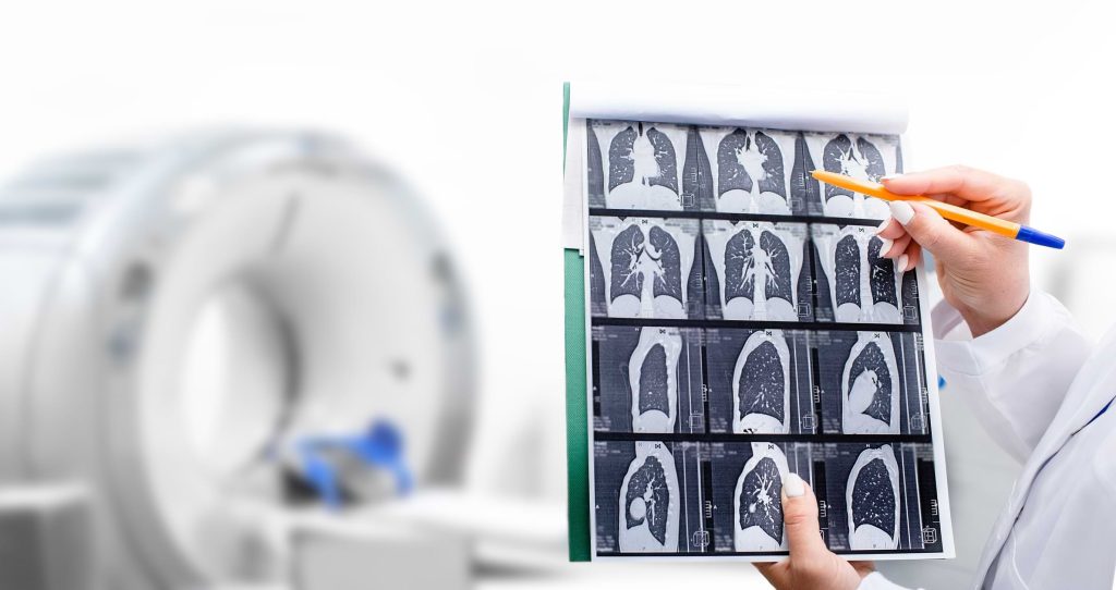
Computed Tomography (CT scans) go a step further by combining multiple X-ray images to create cross-sectional views of organs and tissues. CT scans are faster and offer more detailed imagery, making them vital in emergency settings and complex diagnoses.
CT applications at NIRS:
- Early detection of cancers and tumors.
- Diagnosing brain injuries and strokes.
- Evaluating organ damage in trauma patients.
- Planning surgical or radiotherapy treatments.
MRI and Ultrasound
Magnetic Resonance Imaging (MRI) uses strong magnetic fields and radio waves to produce highly detailed images of internal soft tissues without radiation. MRI is crucial for diagnosing neurological, musculoskeletal, and cardiovascular conditions. NIRS MRI scanners are equipped with advanced features that ensure clarity, speed, and patient comfort.
Common uses of MRI at NIRS:
- Detecting brain tumors, multiple sclerosis, and spinal cord anomalies.
- Diagnosing ligament tears, herniated discs, and joint injuries.
- Visualizing heart structure and function.
Ultrasound imaging (or sonography) uses high-frequency sound waves to create real-time images of internal organs. It is widely used because it is safe, portable, and free from radiation.
NIRS ultrasound services are used for:
- Monitoring pregnancies and fetal development.
- Evaluating abdominal organs like liver, kidneys, and pancreas.
- Detecting gallstones, cysts, and fluid buildup.
- Guiding biopsies and drainage procedures.
PET Scans and Nuclear Medicine
Positron Emission Tomography (PET) scans are a powerful imaging modality used to observe metabolic processes in the body. By using small amounts of radioactive tracers, PET scans can detect how tissues and organs are functioning rather than just their structure.
At NIRS, PET scans are essential for:
- Diagnosing and staging cancers.
- Monitoring tumor response to treatment.
- Evaluating brain disorders such as Alzheimer’s disease.
- Assessing heart muscle viability in cardiac patients.
Nuclear medicine imaging also involves the use of radiopharmaceuticals to examine organ function and structure. It offers functional information that complements structural scans like CT or MRI.
Nuclear imaging at NIRS includes:
- Thyroid scans to detect hyperthyroidism or nodules.
- Bone scans to find infections, fractures, or metastatic disease.
- Renal scans to evaluate kidney function and obstruction.
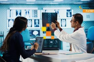
These diagnostic imaging techniques form the backbone of clinical decision-making at NIRS. The institute’s commitment to using the most advanced imaging systems, along with expert radiologists and technologists, ensures accuracy, early detection, and a safer diagnostic journey for every patient.
Case Studies and Real-Life Examples
Real-life applications of diagnostic imaging demonstrate how early detection and accurate diagnosis can change lives. At NIRS (Northern Institute of Radiological Sciences), numerous success stories highlight the pivotal role imaging plays in saving lives and improving outcomes. Here are two compelling examples:
Cancer Detection Through Imaging
Case Study: Early Lung Cancer Diagnosis in a 52-Year-Old Smoker
Mr. A, a 52-year-old chronic smoker, visited NIRS for a routine chest X-ray due to persistent coughing. While the symptoms seemed minor, the digital X-ray revealed a subtle mass in the right upper lobe of his lung. A follow-up CT scan confirmed a small, irregular lesion suggestive of malignancy. NIRS’s radiologist team recommended a PET scan, which showed increased metabolic activity—highly indicative of cancer.
Within days:
- A biopsy was guided by CT imaging.
- Pathology confirmed stage 1 non-small cell lung carcinoma.
- Early detection allowed for minimally invasive surgery without chemotherapy.
Outcome: The patient recovered completely and remains cancer-free after 3 years. Without imaging, this cancer might have progressed silently to a later, untreatable stage.
Cardiovascular Disease Diagnosis
Case Study: Undiagnosed Coronary Artery Disease in a 45-Year-Old Male
Mr. B, a seemingly healthy man with no prior history of heart issues, experienced occasional chest discomfort. He visited NIRS on the advice of his general physician. A non-invasive cardiac CT angiography revealed:
- Significant narrowing in the left anterior descending (LAD) artery.
- No visible symptoms, but a clear blockage risk for future heart attack.
NIRS’s expert cardiovascular imaging team collaborated with cardiologists:
- MRI of the heart confirmed early-stage ischemia.
- A nuclear medicine stress test further evaluated functional heart performance.
- Based on this imaging, a stent procedure was scheduled preemptively.
Outcome: Mr. B avoided a major cardiac event. Imaging not only diagnosed the issue before symptoms escalated but also prevented a possible life-threatening heart attack.
These cases reflect how early detection through advanced diagnostic imaging at NIRS has led to timely treatment, improved prognosis, and better quality of life for patients. Whether it’s catching cancer at stage one or preventing a silent heart attack, diagnostic imaging can be life-saving when combined with expertise and cutting-edge technology.
Future of Diagnostic Imaging in Early Detection
As technology continues to evolve, the future of diagnostic imaging holds incredible promise, particularly in enhancing the early detection of diseases. At NIRS (Northern Institute of Radiological Sciences), we remain at the forefront of adopting innovations that make imaging faster, more accurate, and patient-centered.
1. Integration of Artificial Intelligence (AI) and Machine Learning
AI is revolutionizing medical imaging. At NIRS, we’re beginning to implement AI-powered algorithms that can:
- Analyze imaging scans in real time
- Detect minute anomalies that might be missed by the human eye
- Reduce diagnostic errors and improve workflow efficiency
This ensures faster and more reliable early diagnosis, particularly in complex cases like brain tumors or microvascular heart diseases.
2. Advances in Molecular Imaging
Molecular imaging, such as hybrid PET/MRI, enables clinicians at NIRS to visualize not just structures, but also biological processes at the cellular level. This future-facing approach means:
- Earlier detection of cancers before structural changes occur
- Tracking biochemical changes during treatment
- Personalized medicine strategies
3. Development of Low-Dose Imaging Technology
NIRS prioritizes patient safety, which is why we are exploring low-dose CT and radiation-free imaging techniques such as advanced ultrasound and MRI technologies. These are especially useful for:
- Pediatric and prenatal imaging
- Repetitive scans needed in chronic conditions
4. Wearable Imaging and Portable Devices
The future may see wearable ultrasound devices and mobile diagnostic scanners. While still in early development, NIRS is monitoring such trends closely for their potential to:
- Enable home-based disease monitoring
- Increase access in remote or underserved regions
5. Predictive and Preventive Diagnostics
By combining genetic data, electronic health records, and longitudinal imaging data, NIRS aims to move from reactive healthcare to predictive diagnostics, catching diseases long before symptoms arise.
Challenges and Limitations in Diagnostic Imaging
While diagnostic imaging has transformed healthcare, it’s important to recognize and address the challenges and limitations that come with it. At NIRS, we actively work to minimize these barriers to provide patients with accurate, timely, and safe imaging solutions.
1. Radiation Exposure Risks
Despite many advancements, ionizing radiation remains a concern in imaging techniques like X-rays, CT scans, and PET scans. At NIRS:
- We follow ALARA (As Low As Reasonably Achievable) principles
- Use shielding techniques and low-dose protocols
- Recommend imaging judiciously, especially in vulnerable populations like children and pregnant women
2. Limited Access in Rural or Low-Income Areas
High-end diagnostic imaging devices such as MRI and PET-CT are expensive and not always available in remote regions. NIRS addresses this by:
- Offering community-based imaging programs
- Partnering with local clinics for tele-radiology services
- Expanding outreach through mobile imaging units
3. Cost and Insurance Limitations
Advanced imaging procedures can be costly, especially when not covered fully by insurance. At NIRS, we:
- Maintain transparent pricing
- Offer flexible payment plans
- Work closely with insurers to reduce the financial burden on patients
4. Interpretation Errors and Human Bias
No system is infallible. Misinterpretation or oversight in scan reports can lead to delayed diagnoses. To address this, NIRS:
- Employs board-certified radiologists with sub-specialties
- Uses AI tools as a second opinion system
- Conducts regular peer reviews and quality control
5. Overdiagnosis and Incidental Findings
Some scans may reveal incidental findings (e.g., benign nodules), leading to unnecessary stress or procedures. NIRS physicians:
- Carefully evaluate clinical relevance
- Communicate findings clearly with patients and referring doctors
- Follow evidence-based guidelines to avoid overtreatment
By acknowledging these limitations and continuously improving, NIRS upholds its commitment to patient-centered diagnostic care. We believe that with innovation, ethical practice, and accessibility, diagnostic imaging can reach its full potential in revolutionizing early detection and prevention of disease.
Conclusion
Diagnostic imaging has undeniably revolutionized the medical world by enabling healthcare providers to detect diseases early, often before symptoms appear. From X-rays and MRIs to cutting-edge PET scans, these tools allow for timely diagnosis, improved treatment outcomes, and better patient survival rates. At the heart of this transformative capability lies institutions like NIRS (Northern Institute of Radiological Sciences), which prioritize advanced diagnostic technology, skilled radiologists, and a patient-first approach. In an era where early detection can make the difference between life and death, choosing a center like NIRS ensures you’re in capable and caring hands.
Why Choose NIRS for Diagnostic Imaging?
When it comes to accurate, reliable, and early disease detection, NIRS stands out among diagnostic centers. Here’s why patients in Abbottabad and beyond trust NIRS:
- Cutting-Edge Technology: NIRS is equipped with the latest diagnostic imaging machines, including 1.5 Tesla MRI, multi-slice CT scanners, and digital X-rays that ensure precise results.
- Experienced Radiologists: The institute boasts top-tier radiologists with expertise in detecting complex conditions, ensuring you receive expert interpretations.
- Fast & Accurate Reports: NIRS is known for delivering timely, clear, and detailed reports — critical for quick treatment planning.
- Affordable Services: High-quality diagnostic imaging doesn’t have to be expensive. NIRS offers competitive pricing without compromising quality.
- Patient-Centered Approach: At NIRS, every patient is treated with care, empathy, and respect throughout their diagnostic journey.
Choosing NIRS means choosing reliability, speed, and accuracy — all essential elements in today’s healthcare landscape.
Frequently Asked Questions (FAQs)
Q1. What is diagnostic imaging and why is it important?
A: Diagnostic imaging refers to non-invasive procedures like X-rays, MRIs, CT scans, and ultrasounds that help doctors visualize the internal structures of the body. It’s crucial for early disease detection and treatment planning.
Q2. Is diagnostic imaging safe?
A: Yes. Techniques like ultrasound and MRI use no radiation, while CT scans and X-rays use minimal doses. NIRS ensures patient safety with every scan, following strict protocols.
Q3. How long does it take to get results at NIRS?
A: Most diagnostic imaging reports are available within 24 to 48 hours at NIRS, depending on the test.
Q4. Do I need a doctor’s referral to get a scan at NIRS?
A: While referrals are helpful, you can consult directly with NIRS’s on-site specialists for most imaging services.
Q5. Can early detection through imaging really improve survival?
A: Absolutely. Diseases like cancer, heart issues, and infections are often treatable when caught early. Diagnostic imaging significantly enhances survival chances by enabling early intervention.
Contact Information
For premium diagnostic imaging services in Abbottabad, contact NIRS (Northern Institute of Radiological Sciences):
- 📞 Phone: +92316 5212015
- 📧 Email: Info@nirs.com.pk
- 🌐 Website: www.nirs.com.pk
- 📍 Location: IDC Small industrial Chowk Mandian Abbottabad Near Ayub Medical Complex
- 💬 Facebook: facebook.com/NIRS
- 📸 Instagram: instagram.com/NIRS
At NIRS, your health comes first — with world-class diagnostic imaging that leads the way in early disease detection.